
- >Scientific productions
- >Thesis
- >Pauline TEIXEIRA thesis defence
Pauline TEIXEIRA thesis defenceSous la direction du Dr Pascale May-Panloup et co-direction du Dr Arnaud Chevrollier
The December 11, 2025
Jeudi 11 décembre 2025 | 09h30 | Amphithéâtre ICO.
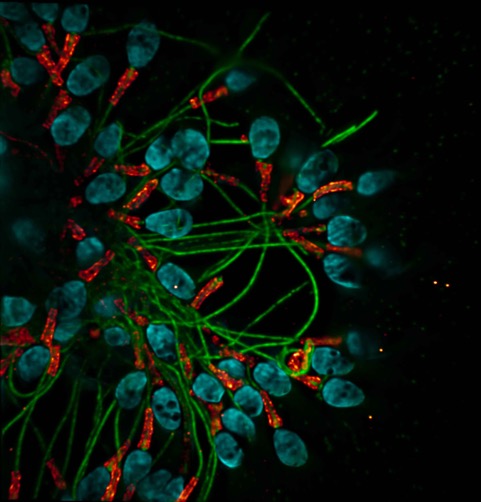
Study of mitochondrial structure and function and their implications in male infertility.
Abstract :
Infertility affects nearly one in six couples and is therefore a major public health issue. Among the factors involved, sperm quality is declining, particularly in Western societies. Sperm is a highly specialised cell that contains half of the genetic material and is equipped with a flagellum that gives it mobility. This mobility requires ATP, which is produced in particular by the mitochondrial membrane. In most tissues, mitochondria are highly dynamic tubular structures that divide, fuse, change shape and move. In spermatozoa, however, mitochondria have a fixed organised structure: they are wrapped around the axoneme in the midpiece of the flagellum.
Various alterations in sperm parameters have been associated with mitochondrial abnormalities. We used high-resolution fluorescence microscopy techniques to study the structure of mitochondria in spermatozoa as well as the location and expression of proteins involved in their function.
We highlight a deregulation of proteins associated with oxidative phosphorylation in the spermatozoa of patients with oligoasthenospermia. We also show the persistence of nucleoids in patients from the same group. Finally, we specify the atypical location of mitochondrial transcription factor A in spermatozoa.
These results provide new insights into the structure-function relationship of mitochondria in spermatozoa and their involvement in alterations in sperm quality.
Keywords : Spermatozoa, Mitochondria, Male infertility, Microscopy.
Etude de la structure et de la fonction mitochondriale et leurs implications dans l’infertilité masculine.
Résumé :
L’infertilité concerne près d'un couple sur six et constitue ainsi un enjeu majeur de santé publique. Parmi les facteurs en cause, la qualité du sperme est en déclin, en particulier dans les sociétés occidentales. Le spermatozoïde est une cellule hautement spécialisée, que contient un hémi-patrimoine génétique et qui est munie d’un flagelle qui lui confère sa mobilité. Cette mobilité nécessite de l’ATP produit notamment par la gaine mitochondriale. Dans la plupart des tissus, les mitochondries sont des structures tubulaires très dynamiques qui se divisent, fusionnent, changent de forme et se déplacent. Dans les spermatozoïdes, en revanche, les mitochondries ont une structure organisée fixe : elles sont enroulées autour de l’axonème dans la pièce intermédiaire du flagelle.
Diverses altérations des paramètres spermatiques ont été associées à des anomalies mitochondriales. Nous avons utilisé des techniques de microscopie fluorescente à haute-résolution pour étudier la structure des mitochondries dans le spermatozoïde ainsi que la localisation et l’expression de protéines nécessaires à leur fonctionnement.
Nous avons mis en évidence une dérégulation des protéines associées aux phosphorylations oxydatives dans les spermatozoïdes de patients oligoasthénospermiques. Nous avons montré aussi la persistance de nucléoïdes chez des patients du même groupe. Enfin, nous avons précisé la localisation atypique du facteur de transcription mitochondrial A dans les spermatozoïdes.
Ces résultats apportent de nouveaux éléments sur la relation structure-fonction des mitochondries dans les spermatozoïdes et leurs implications dans les altérations de la qualité spermatique.
Mots clés : Spermatozoïde, Mitochondrie, Infertilité masculine, Microscopie.
